Filter results by Topics

Your search for all content returned 252 results

Save search You must be logged in as an individual save a search. Log-in/register
Book
Endocrine Physiology, 6th Edition

by Patricia E. Molina

Master the aspects of endocrine physiology required in clinical medicine and to ace the USMLE

Endocrine Physiology delivers unmatched coverage of the fundamental concepts of hormone biological actions - providing the foundation you need to understand the physiologic mechanisms involved in neuroendocrine regulation of organ function.

This updated edition has been revised for greater clarity and understanding. Each chapter opens with a short description of the functional anatomy of the organ, highlighting important features pertaining to circulation, location, or cellular composition that have a direct effect on endocrine function. Newly added annotated illustrations highlight principle concepts in each chapter.

Emphasizing must-know principles, Endocrine Physiology is the single-best endocrine review available for the USMLE Step 1.

This sixth edition features:

• An informative first chapter describing the organization of the endocrine system, as well as general concepts of hormone production and release, transport and metabolic rate, and cellular mechanisms of action

• Case studies that show how to apply principles to real-world clinical situations

• Bulleted objectives, key concepts, study questions with expanded answers, suggested readings, and diagrams encapsulating key concepts

• Pedagogical instruction throughout

Book
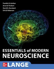
Essentials of Modern Neuroscience, 1st Edition

by Franklin R. Amthor, Anne B. Theibert, David G. Standaert, Erik D. Roberson

Created by an expert team of neuroscience educators, this comprehensive guide delivers the knowledge and insight you need to build your understanding of neuroscience—quickly and easily. Divided into two parts, the guide offers a thorough treatment of the basic science of the anatomy and function of the nervous system, as well as an extended treatment of nervous system disorders and therapeutics.

Packed with 500 color illustrations, Essentials of Modern Neuroscience provides both clinical content and numerous cases in an engaging, simple-to-understand style. It includes the strong pedagogy that makes LANGE basic science titles so popular and provides chapter-opening Learning Objectives, bulleted chapter summaries, and application boxes.

Features:

• Covers both basic science and clinical cases for full mastery of the topic

• Organized to mirror the way medical schools teach neuroscience

• Presents information in a way that fosters maximum retention

• Unique chapters cover addiction, affective disorders, and neurologic diseases

• Outstanding art program, where readers can visually learn from the images and legends

Book
Greenspan's Basic & Clinical Endocrinology, 10th Edition

by David G. Gardner, Dolores Shoback

A full-color guide to the entire field of clinical endocrinology and its scientific underpinnings—updated with the latest breakthroughs and developments.

A Doody's Core Title for 2021!

Greenspan's Basic & Clinical Endocrinology, Tenth Edition delivers a succinct, leading-edge overview of the underlying molecular biology of the endocrine system and the latest perspectives on the diagnosis and treatment of specific diseases and disorders. Featuring an enhanced design that includes hundreds of full-color illustrations and clinical photographs, Greenspan's is a true must-have during traditional or integrated courses in endocrinology, endocrinology rotation, or exam prep in internal medicine and endocrinology, as well as a reference for disease management.

Greenspan's provides clinically relevant coverage of metabolic bone disease, pancreatic hormones and diabetes mellitus, hypoglycemia, obesity, and many other diseases and disorders. Supporting this important material is a handy appendix of normal hormone reference ranges across the lifespan.

Here's why Greenspan's is an essential tool for learning how to manage endocrine patients:

• The Tenth Edition is enhanced by updated content throughout each chapter

• NEW CHAPTERS on Transgender Endocrinology and Disorders of Sexual Determination and Differentiation

• Important chapter on Evidence-Based Endocrinology and Clinical Epidemiology

• Concise, balanced coverage of both scientific and clinical principles that guide patient management

• The best source for current concepts in endocrine pathophysiology to aid clinical decision making

• The most practical, current insights into diagnostic testing

• More than 270 full-color illustrations and clinical photographs

If you are in need of a well-illustrated, completely up-to-date guide to the entire field of clinical endocrinology, this trusted classic belongs on your desk or computer.

Book
Harper's Illustrated Biochemistry, 32nd Edition

by Peter J. Kennelly, Kathleen M. Botham, Owen P. McGuinness, Victor W. Rodwell, P. Anthony Weil

Gain a thorough understanding of the principles of biochemistry as they relate to clinical medicine

The thirty-second edition of Harper's Illustrated Biochemistry combines top-quality, full-color illustrations with authoritative, integrated coverage of biochemical disease and clinical information. Featuring numerous medically relevant examples, this respected text presents a clear, succinct review of the fundamentals that every student must understand in order to succeed in medical school.

All 58 chapters help you understand the medical relevance of biochemistry.

• Full-color presentation with 600+ illustrations

• Chapters have been updated to reflect the latest information

• Case studies emphasize the clinical relevance of biochemistry

• Review questions follow each of the 11 sections

• Boxed objectives define the goals of each chapter

• Tables encapsulate important information

• Each chapter contains a section on biomedical importance and a summary of the topics covered

Applauded by medical students for its current and engaging style, Harper's Illustrated Biochemistry is an essential for USMLE review and the single best reference for learning the clinical relevance of any biochemistry topic.

Book
Junqueira's Basic Histology: Text and Atlas, 17th Edition

by Anthony L. Mescher

The text that has defined histology for generations–concise, clear, beautifully illustrated, and updated with new content and more Q&A!

For more than 50 years, Junqueira's Basic Histology has been considered the hands-down best overview of human tissue structure and function available. Accessible yet comprehensive, this trusted classic provides everything you need to know about basic cell biology and histology, integrating the material with that of biochemistry, immunology, endocrinology, and physiology. With coverage of all tissues, every organ system, organs, bone and cartilage, blood, skin, and more, Junqueira's is a valuable foundation for subsequent studies in pathology.

Formatted to optimize the learning process, Junqueira's is filled with clear explanations, art, and micrographs to clarify key concepts. This is an essential resource for students of medicine and other health-related professions, as well as for advanced undergraduate courses in tissue biology.

Features:

• Self-test questions in every chapter

• New: 550 Self-assessment Q&As – more than twice that of the previous edition!

• Key points and summary tables highlighting key content

• Clinical correlations for each topic

• Illustrations depicting key aspects of cell biology and histology

• Electron and light micrographs that deliver a definitive atlas of cell, tissue, and organ structures

• Valuable Appendix that explains light microscopy stains

• Lab manual guides readers to see and identify in actual tissue specimens all cells and tissues understudy

• New: PowerPoint slides with illustrations and micrographs via AccessMedicine

Book
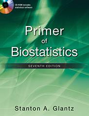
Primer of Biostatistics, 7th Edition

by Stanton A. Glantz

A concise, engagingly written introduction to understanding statistics as they apply to medicine and the life sciences

CD-ROM performs 30 statistical tests

Don't be afraid of biostatistics anymore! Primer of Biostatistics, 7th Edition demystifies this challenging topic in an interesting and enjoyable manner that assumes no prior knowledge of the subject. Faster than you thought possible, you'll understand test selection and be able to evaluate biomedical statistics critically, knowledgeably, and confidently.

With Primer of Biostatistics, you'll start with the basics, including analysis of variance and the t test, then advance to multiple comparison testing, contingency tables, regression, and more. Illustrative examples and challenging problems, culled from the recent biomedical literature, highlight the discussions throughout and help to foster a more intuitive approach to biostatistics.

The companion CD-ROM contains everything you need to run thirty statistical tests of your own data. Review questions and summaries in each chapter facilitate the learning process and help you gauge your comprehension. By combining whimsical studies of Martians and other planetary residents with actual papers from the biomedical literature, the author makes the subject fun and engaging.

Coverage includes:

• How to summarize data

• How to test for differences between groups

• The t test

• How to analyze rates and proportions

• What does "not significant" really mean?

• Confidence intervals

• How to test for trends

• Experiments when each subject receives more than one treatment

• Alternatives to analysis of variance and the t test based on ranks

• How to analyze survival data

Book
Schaum’s Outline of Biochemistry, 3rd Edition

by Philip W. Kuchel, Simon Easterbrook-Smith, Vanessa Gysbers, J. Mitchell Guss

Confused about biochemistry? Problem solved. This easy-to-follow outline of the typical one-year course in biochemistry helps you grasp course content and prepare for exams. New chapters cover dramatic recent discoveries about how cells respond to changes in their environment, as well as new developments in genetic engineering and the human genome project. Ideal not just for science majors, but also for students in the health sciences (medicine, dentistry, vet science, nursing, etc).

Book
Sherris & Ryan's Medical Microbiology, 8th Edition

by Kenneth J. Ryan

The most dynamic, comprehensive, and student-friendly text on the nature of microorganisms and the fascinating processes they emply in producing infection and disease.

For more than a quarter-of-a-century, this renowned text has helped readers develop a solid grasp of the significance of etiologic agents, the pathogenic processes, epidemiology, and the basis of therapy for infectious diseases. Now, with a NEW four-color design, the book is shorter and more assessable for students! Outstanding pedagogical elements are carried throughout this edition including:

• Over 400 outstanding images with hundreds of tables and illustrations

• Detailed legends under the art so the reader can better understand what's occurring within the illustration, without having to flip back to the text

• Clinical Cases with USMLE Style Questions

• Margin Notes identifying the "high-yield" must know content in each chapter

• Bulleted Summaries that conclude each chapter

Sherris & Ryan's Medical Microbiology, Eighth Edition is divided into five parts:

Part I opens with a chapter that explains the nature of infection and the infectious agents at the level of a general reader. The following four chapters give more detail on the immunologic, diagnostic, and epidemiologic nature of infection with minimal detail about the agents themselves.

Parts II through V form the core of the text with chapters on the major viral, bacterial, fungal, and parasitic diseases, and each begins with its own chapters on basic biology, pathogenesis, and antimicrobial agents.

Features and Learning Aids:

• 57 chapters that simply and clearly describe the strains of viruses, bacteria, fungi, and parasites that can bring about infectious diseases (plus one online only chapter)

• Explanations of host-parasite relationship, dynamics of infection, and host response

• A clinical case with USMLE-style questions concludes each chapter on the major viral, bacterial, fungal, and parasitic diseases

• Numerous full-color photographs, tables, and illustrations

• Clinical Capsules cover the essence of the disease(s) caused by major pathogens

• Chapter-ending case questions PLUS a collection of 100 practice questions

• Innovative study aids including boxed narrative Overviews that open each disease-oriented chapter or major section, highlighted Margin Notes pointing out high-yield material for USMLE Step 1 preparation, bulleted lists of Key Conclusions at the end of each major section, a THINK - APPLY feature that randomly inserts thought-provoking questions into the body of the text, and more.

• A set of tables that presents the microbes in context of the clinical infections they produce

Book Chapter
28. Actinomyces and Nocardia

28. Actinomyces and Nocardia

OVERVIEW

Actinomycosis is a chronic inflammatory condition originating in the tissues adjacent to mucosal surfaces caused by anaerobic Gram-positive branching bacilli of the genus Actinomyces that are present in the microbiota of the alimentary tract. Disease occurs when minor trauma displaces these bacteria below the mucosal barrier. The lesions follow a slow burrowing course with considerable induration and draining sinuses, eventually opening through the skin. The exact nature depends on the organs and structures involved.

Actinomyces are typically elongated Gram-positive rods that branch at acute angles (Figure 28–1). They are Gram-positive bacilli that grow slowly (4-10 days) under microaerophilic or strictly anaerobic conditions. In pus and tissues, the most characteristic form is the sulfur granule (Figure 28–2). This yellow-orange granule, named for its gross resemblance to a grain of sulfur, is a microcolony of intertwined branching Actinomyces filaments solidified with elements of tissue exudate.

Figure 28–1 Actinomyces. Note the angular branching of the Gram-positive bacilli. (Reproduced with permission from Willey JM: Prescott, Harley, & Klein’s Microbiology, 7th ed. New York, NY: McGraw Hill; 2008.)
ryan8_ch28_f001
Figure 28–2 Sulfur granule. The mass is a microcolony of bacteria Gram-positive bacteria and tissue elements. The branching is clearly seen only at the edge. (Reproduced with permission from Connor DH, Chandler FW, Schwartz DQ, et al: Pathology of Infectious Diseases. Stamford CT: Appleton & Lange; 1997.)
ryan8_ch28_f002

❋ Anaerobic branching Gram-positive rods

Species of Actinomyces are distinguished on the basis of biochemical reactions, cultural features, and cell wall composition. Most human actinomycosis is caused by Actinomyces israelii, but other species have been isolated from typical actinomycotic lesions. Another group of Actinomyces species have been associated with dental and periodontal infections (see Chapter 41).

❋ Most infections A israelii

Actinomyces are normal inhabitants of some areas of the gastrointestinal tract of humans and animals from the oropharynx to the lower bowel. These species are highly adapted to mucosal surfaces and do not produce disease unless they transgress the epithelial barrier under conditions that produce a sufficiently low oxygen tension for their multiplication (Figure 28–3). Such conditions usually involve mechanical disruption of the mucosa with necrosis of deeper, normally sterile tissues (eg, following tooth extraction). Once initiated, growth occurs in microcolonies in the tissues and extends without regard to anatomic boundaries. The lesion is composed of inflammatory sinuses, which ultimately discharge to the surface. As the lesion enlarges, it becomes firm and indurated. Sulfur granules are present within the pus but are not numerous. Free Actinomyces or small branching units are rarely seen, although contaminating Gram-negative rods are common. As with other anaerobic infections, most cases are polymicrobial involving other flora from the mucosal site of origin including other Actinomyces species.

Figure 28–3 Actinomycosis and Nocardiosis. (Right) Actinomyces are members of the normal flora throughout the alimentary tract. Minor trauma allows access to tissues where they create burrowing abscesses that may break through to the surface. (Left) Nocardia is present in the soil, where it may be either inhaled to produce a pneumonia or traumatically injected to produce cutaneous pustules and lymphadenitis.
ryan8_ch28_f003

❋ Microbiota in gastrointestinal tract

❋ Displacement into tissues

Sinus tracts with sulfur granules

Human cases of actinomycosis provide little evidence of immunity to Actinomyces. Once established, infections typically become chronic and resolve only with the aid of antimicrobial therapy. Antibodies can be detected in the course of infection, but seem to reflect the antigenic stimulation of the ongoing infection rather than immunity. Infections with Actinomyces are endogenous, and case-to-case transmission does not appear to occur.

Little evidence of immunity

Actinomycosis exists in several forms that differ according to the original site and circumstances of tissue invasion. Infection of the cervicofacial area, the most common site of actinomycosis (Figure 28–4), is usually related to poor dental hygiene, tooth extraction, or some other trauma to the mouth or jaw. Lesions in the submandibular region and the angle of the jaw give the face a swollen, indurated appearance.

Figure 28–4 Cervicofacial actinomycosis. The classic “lumpy jaw” is shown with draining sinuses at the angle of the jaw. The lesion would be very firm on palpation. (Reproduced with permission from Connor DH, Chandler FW, Schwartz DQ, et al: Pathology of Infectious Diseases. Stamford CT: Appleton & Lange; 1997.)
ryan8_ch28_f004

❋ Linked to poor dental hygiene

Thoracic and abdominal actinomycosis are rare and follow aspiration or traumatic (including surgical) introduction of infected material leading to erosion through the pleura, chest, or abdominal wall. Diagnosis is usually delayed because only vague or nonspecific symptoms are produced until a vital organ is eroded or obstructed. The firm, fibrous masses are often initially mistaken for a malignancy. Pelvic involvement as an extension from other sites also occurs occasionally. It is particularly difficult to distinguish from other inflammatory conditions or malignancies. A more localized chronic endometritis, due to Actinomyces, is associated with the use of intrauterine contraceptive devices.

Surgery, trauma, intrauterine devices provide access

A clinical diagnosis of actinomycosis is based on the nature of the lesion, the slowly progressive course, and a history of trauma or of a condition predisposing to mucosal invasion by Actinomyces. The etiologic diagnosis can be difficult to establish with certainty. Although the lesions may be extensive, the organisms in pus may be few and concentrated in sulfur granule microcolonies deep in the indurated tissue. The diagnosis is further complicated by heavy colonization of the moist draining sinuses with other bacteria, usually Gram-negative rods. This contamination not only causes confusion regarding the etiology but interferes with isolation of the slow-growing anaerobic Actinomyces. Material for direct smear and culture should include as much pus as possible to increase the chance of collecting the diagnostic sulfur granules.

Sinus drainage contains few Actinomyces

Sulfur granules crushed and stained show a dense, Gram-positive center with individual branching rods at the periphery (Figure 28–2). Granules should also be selected for culture, because material randomly taken from a draining sinus usually grows only superficial contaminants. Culture media and techniques are the same as those used for other anaerobes. Incubation must be prolonged because some strains require 7 days or more to appear. Identification requires a variety of biochemical tests to differentiate Actinomyces from Propionibacteria, which may show a tendency to form short branches.

❋ Gram stains show branching rods

Anaerobic culture required

Biopsies for culture and histopathology are useful, but it may be necessary to examine many sections and pieces of tissue before sulfur granule colonies of Actinomyces are found. The morphology of the sulfur granule in tissue is quite characteristic with routine hematoxylin and eosin (H&E) or histologic Gram staining. With the histologic H&E stain, the edge of the granule shows amorphous eosinophilic “clubs” formed from the tissue elements and containing the branching actinomycotic filaments.

Biopsy shows clubbed lesions

Penicillin G is the treatment of choice for actinomycosis, although a number of other antimicrobics (ampicillin, doxycycline, erythromycin, and clindamycin) are active in vitro and have shown clinical effectiveness. Metronidazole is not active. High doses of penicillin must be used and therapy prolonged for up to 6 weeks or longer before any response is seen. The initial treatment course is usually followed with an oral penicillin for 6 to 12 months. Although slow, response to therapy is often striking given the degree of fibrosis and deformity caused by the infection. Because detection of the causative organism is difficult, many patients are treated empirically as a therapeutic trial based on clinical findings alone.

Penicillin is effective

KEY CONCLUSIONS

  • Anaerobic branching Gram-positive rods grow in microcolonies called sulfur granules.

  • Displacement from microbiota habitat across oropharyngeal or intestinal mucosa leads to burrowing lesions.

  • Culture diagnosis from draining sinuses complicated by contaminating bacteria.

  • Penicillin and other β-lactams are effective treatment.

Book Chapter
6. Adipose Tissue

6. Adipose Tissue

Specialized for relatively long-term energy storage, adipocytes of white adipose tissue become spherical when isolated but are polyhedral when closely packed in situ. When completely developed, a white adipocyte is very large, between 50 and 150 μm in diameter, and contains a single huge droplet of lipid filling almost the entire cell. Having a single large droplet of triglycerides, white adipocytes are also called unilocular (Figure 6–1). Sometimes described as having a signet-ring appearance, an adipocyte’s single lipid droplet displaces most cytoplasm and flattens the nucleus against the cell membrane (Figure 6–1d). Because lipid undergoes removal from cells by xylene or other solvents used in routine histological techniques, unilocular adipocytes often appear empty in standard light microscopy. This membrane and the thin rim of cytoplasm that remains after such loss of the stored lipid frequently shrinks, collapses, or ruptures, distorting cell and tissue structure.

Figure 6–1 White adipose tissue. White or unilocular adipose tissue is commonly seen in sections of many human organs. (a) Large white adipocytes (A) are seen in the connective tissue associated with small blood vessels. The fat cells are empty because lipid was dissolved away in slide preparation. Nuclei at the cell membranes are visible in some of the fat cells. (×100; H&E) (b) Large (empty) adipocytes predominate in this typical white adipose tissue, which shows only a small portion of microvasculature. In a single histologic section, nuclei of most very large adipocytes are not included. (×100; H&E) (c) Tissue was fixed here with osmium tetroxide, which preserves lipid (L) and stains it black. Many adipocytes in this slide retain at least part of their large lipid droplets. (×440; Osmium tetroxide) (d) In this specimen from a young mammal the smaller adipocytes marked with asterisks are not unilocular, having many lipid droplets of various sizes. Such cells in white fat represent those in which differentiation is incomplete as well as a small subpopulation of beige cells with brown fat-forming potential. The eccentric nuclei of the unilocular cells are indicated by arrowheads. (×200; PT)
mescher17_ch6_f001

MEDICAL APPLICATION

Unilocular adipocytes can generate relatively common benign tumors called lipomas, although malignant adipose tumors (liposarcomas) occur infrequently. Fetal lipomas of brown fat are sometimes called hibernomas.

Most cytoplasmic organelles in a white adipocyte localize near the peripheral nucleus, including mitochondria, a small Golgi apparatus, a few cisternae of RER, and free polyribosomes. The thin, submembranous layer of cytoplasm surrounding the lipid droplet contains cisternae of smooth ER (SER) and pinocytotic vesicles. Transmission electron microscopy (TEM) reveals a great abundance of caveolae in the cell membranes of most adipocytes, especially immature cells, and numerous minute lipid droplets in addition to the large droplet. In this cell type caveolae play important roles in lipid trafficking and formation of the large triglyceride storage droplet.

As shown in Figure 6–1, partitions of connective tissue containing a vascular bed and a nerve network subdivide white fat into incomplete lobules. Fibroblasts, macrophages, and other cells typically comprise about half the total cell number in white adipose tissue. Reticular fibers form a fine interwoven network supporting individual fat cells and binding them together. The microvasculature between adipocytes may not always be apparent in tissue sections.

The distribution of white adipose tissue changes significantly through childhood and adult life, partly regulated by sex hormones controlling adipose deposition in the breasts and thighs. The color of freshly dissected white adipose tissue depends on the diet, varying from white to yellow with increasing amounts of carotenoid dissolved in the lipid.

Storage & Mobilization of Lipids

White adipocytes can store triglycerides derived from three sources:

  • Dietary fats brought to the cells via the circulation as chylomicrons,

  • Lipids synthesized in the liver and transported in blood with very low-density lipoproteins (VLDLs), and

  • Free fatty acids and glycerol synthesized by the adipocytes.

Chylomicrons (Gr. chylos, juice + micros, small) represent particles of variable size, up to 1200 nm in diameter, formed from ingested lipids in epithelial cells lining the small intestine and transported in the blood and lymph. They consist of a core containing mainly triglycerides, surrounded by a stabilizing monolayer of phospholipids, cholesterol, and several apolipoproteins.

VLDLs appear as much smaller complexes (30–80 nm, providing a greater surface-to-volume ratio) of similar lipid and protein composition to chylomicrons, but undergo synthesis and release in liver cells. Clinical tests for circulating levels of lipoproteins routinely measure blood lipids after fasting to allow depletion of chylomicrons. Varying levels of apoproteins and triglycerides in the complexes allow their categorization according to density, from VLDL to high-density lipoprotein (HDL).

In adipose tissue both chylomicrons and VLDLs become hydrolyzed at the luminal surfaces of blood capillaries by lipoprotein lipase, an enzyme synthesized by the adipocytes and transferred to the capillary cell membrane (Figure 6–2). Free fatty acids then enter the adipocytes by both active transport and diffusion. Within the adipocytes the fatty acids combine with glycerol phosphate, supplied by glucose metabolism, to again form triglycerides, which then get deposited in the growing lipid droplet. Insulin stimulates glucose uptake by adipocytes and accelerates its conversion into triglycerides, and the production of lipoprotein lipase.

Figure 6–2 Lipid storage and mobilization from adipocytes. Triglycerides are transported by blood and lymph from the intestine and liver in lipoprotein complexes known as chylomicrons (Chylo) and VLDLs. In the capillary endothelial cells of adipose tissue, these complexes are partly broken down by lipoprotein lipase, releasing free fatty acids and glycerol. The free fatty acids diffuse from the capillary into the adipocyte, where they are reesterified to glycerol phosphate, forming triglycerides that are stored in the lipid droplet until needed. Norepinephrine from nerve endings stimulates the cyclic AMP (cAMP) system, which activates hormone-sensitive lipase to hydrolyze the stored triglycerides to free fatty acids and glycerol. These substances diffuse into the capillary, where the fatty acids bind albumin for transport throughout the body for use as an energy source. Abundant caveolae in the adipocyte plasmalemma are rich in cholesterol and other lipids and appear to mediate endocytosis of fatty acids necessary for growth of the lipid storage droplet.
mescher17_ch6_f002

Upon adipocyte stimulation by nerves or various hormones, stored lipids become mobilized and cells release fatty acids and glycerol. Norepinephrine released in the adrenal gland and by postganglionic sympathetic nerves in adipose tissue activates a hormone-sensitive lipase that breaks down triglycerides at the surface of the stored lipid droplets (Figure 6–2). Growth hormone (GH) from the pituitary gland also stimulates this lipase activity. The free fatty acids diffuse across the membranes of the adipocyte and the capillary endothelium and bind the protein albumin in blood for transport throughout the body. The more water-soluble glycerol remains free in blood for uptake in the liver. Insulin inhibits the hormone-sensitive lipase, reducing fatty acid release, and stimulates enzymes for lipid synthesis. Besides insulin and GH, other peptide hormones also cooperate in regulating lipid synthesis and mobilization in adipocytes.

Hormonal activity of white adipocytes themselves includes production of the 16-kDa polypeptide hormone leptin (Gr. leptos, thin), a “satiety factor” with target cells in the hypothalamus, other brain regions, and peripheral organs, which helps regulate the appetite under normal conditions and participates in regulating the formation of new adipose tissue.

MEDICAL APPLICATION

Leptin was discovered and is well studied in genetically obese mice, but such studies have not yet led to new treatments for human obesity. In most obese humans, adipocytes produce adequate or excess quantities of leptin, but target cells are not responsive due apparently to insufficient or defective receptors or post-receptor signal transduction.

Although white adipose tissue associated with different organs appears histologically similar, differences in gene expression have been noted between visceral deposits (in the abdomen) and subcutaneous deposits of white fat. Such differences may have importance for medical risks of obesity; it is well established that increased visceral adipose tissue raises the risk of diabetes and cardiovascular disease, whereas increased subcutaneous fat does not. The release of visceral fat products directly to the portal circulation of the liver may also influence the medical relevance of this form of obesity.

In response to body needs, lipids undergo mobilization rather uniformly from white adipocytes in all parts of the body, although adipose tissue in the palms, soles, and fat pads behind the eyes resists even long periods of starvation. During starvation, adipocytes can lose nearly all their fat and become polyhedral or spindle-shaped cells with only very small lipid droplets.

Histogenesis of White Adipose Tissue

Like other connective tissue, skeletal and muscle cells, adipocytes develop from mesenchymal stem cells. Adipose development first produces preadipocytes, which look rather like larger fibroblasts with cytoplasmic lipid droplets (Figure 6–3). Initially, the droplets of white adipocytes are separate from one another but they soon fuse to form the single large droplet (Figure 6–1).

Figure 6–3 Development of white and brown fat cells. Mesenchymal stem cells differentiate as progenitor cells for all types of connective tissue, including preadipocytes. These are initially of at least two types. Preadipocytes developing within the lateral mesoderm of the embryo produce large number of white adipocytes (forming white adipose tissue) and a smaller number of so-called “beige” adipocytes with cytological features and gene expression patterns of both white and brown adipocytes. White adipocytes are unilocular, with one large lipid droplet occupying most of the cytoplasm. The white adipocyte is usually much larger than that shown here in relation to the other cell types. Brown adipocytes differentiate from another population of preadipocytes located in paraxial embryonic mesoderm and remain multilocular (having many small lipid droplets) with numerous mitochondria (not shown here). Mitochondrial metabolism of lipid in brown adipocytes releases heat rather than ATP. Cells functioning as brown adipocytes can also develop from beige adipocytes during adaptation to cold temperatures.
mescher17_ch6_f003

As shown in Figure 6–3, white adipocytes develop together with a smaller population of cells termed beige adipocytes, which remain within white adipose tissue and have histological and metabolic features generally intermediate between white and brown adipocytes. With adaptation to cold temperatures beige adipocytes change reversibly, forming many more small lipid droplets, adopting a gene expression profile more like that of brown fat and begin to release heat as described below.

MEDICAL APPLICATION

In addition to leptin, white adipose tissue secretes numerous other cytokines and other factors with paracrine and autocrine activity, including many proinflammatory cytokines. It is not clear whether these are produced by adipocytes or other cells of the tissue such as macrophages or fibroblasts. With its increased amounts of white adipose tissue, obesity is characterized by a state of chronic mild inflammation. Proinflammatory factors released from visceral fat are being investigated for links to the inflammation-related disorders associated with obesity, such as diabetes and heart disease.

At birth humans have stores of white adipose tissue, which begin to accumulate by the 14th week of gestation. Both visceral and subcutaneous fat becomes well developed after this time. Proliferation of progenitor cells diminishes by late gestation, and adipose tissue increases mainly by the filling of existing adipocytes until around age 10, followed by a period of new fat cell differentiation that lasts through adolescence. New adipocyte formation occurs around small blood vessels, where undifferentiated mesenchymal cells are most abundant.

Excessive adipose tissue accumulation, or obesity, occurs when nutritional intake exceeds energy expenditure, an increasingly common condition in modern, sedentary lifestyles. Although adipocytes can differentiate from mesenchymal stem cells throughout life, adult-onset obesity mainly involves increasing the size of existing adipocytes (hypertrophy). Childhood obesity, in contrast, often involves increases in both adipocyte size and numbers due to the differentiation of more preadipocytes from mesenchymal cells (hyperplasia). Weight loss after dietary changes results from reductions in adipocyte volume, but not their overall number.

MEDICAL APPLICATION

Adult-onset obesity is very often associated with age-related metabolic changes and may involve reduced activity of the hormone-sensitive lipases of adipocytes, causing less effective fat mobilization out of the cells. The increased number of adipocytes produced during childhood obesity predisposes an individual to obesity in later life. Despite claims of various fad diets, there is no evidence that any particular type of caloric restriction is more effective than others; rather, any intake of calories that is lower than the energy expenditure will result in loss of adipose tissue.